Monday, October 28, 2013

Elevated Arc/Arg 3.1 protein expression in the basolateral amygdala following auditory trace-cued fear conditioning- Chau- 2013


A: Trace     B: Backward     C: Delay


        From the amygdala


This is the first study examining amygdala activation in closer detail following trace fear associative learning. 
The present experiments demonstrate that the amygdala, specifically the BLA, is activated following trace fear conditioning. 
Activation was not found in the CeA or the LA following trace fear conditioning compared to backward-conditioned, delay-conditioned and naïve mice, suggesting that the BLA plays a more critical role in trace fear associative learning than delay fear associative learning. 
Additionally, findings from this study demonstrate that delay-conditioned mice exhibited more Arc expression in the LA compared to any of the other conditioned groups and controls, suggesting that delay fear conditioning taps into an underlying pathway different from trace fear conditioning. 
Together, these findings suggest that the underlying pathway and thus possible neuronal mechanisms for acquisition and subsequent consolidation for trace fear associations are more similar to contextual fear associations than delay fear associations. 

Friday, October 25, 2013

Creating a false memory in the Hippocampus- Tonegawa- 2013

Question:
- Whether the internal presentation (generated by conscious or unconscious recall, dreaming, and imagination) can be combined with external stimuli to generate new memories (?)
- Or in this study, whether artificially activating a previously formed contextual fear memory engram, would create a false fear memory in a shocks-free context  (?)
- Or whether a light-activated contextual memory in the DG or CA1 can serve as a functional CS in fear conditioning(?)

Lessons from the past experiments: a small subpopulation of granule cells in the DG identified as contextual memory- engram cells. Optogenetic stimulation of these cells were enough to bring back the freezing behavior.

Method related notes:
c-fos-tTA transgenic mice were used.
c-Fos gene promotor------> tTA -------> TRE -----> gene of interest expression (?)
They injected AAV encoding TRE-ChR2-mCherry in to the DG or CA1 of c-fos-tTA mice. 


On Dox diet -----> No (ChR2)-mCherry expression in the DG
Optical stimulation of (ChR2)-mCherry expressing DG cells -----> cFos expression from anterior - posterior axis of DG

Experiment design:


Off Dox in ctxA--- On Dox--- Next day fear-conditioned in ctxB while optically re-activating ctxA labeled cells --- 2 days later tested in ctxA or ctxC
If the light-reactivated cells labeled in ctxA can produce a functional CS during fear conditioning in ctxB, then the animals should express a false fear memory by freezing in ctxA, but not in ctxC. *

Q: The degree of overlap of cells activated in ctxA and ctxC?
Off Dox in ctxA--- On Dox right away--- Next day half mice exposed to ctxA, the other half to ctxC--- 1.5h later sacrifice ------> Statically different populations of DG cells in ctxA and ctxC with overlapping cell populations in dorsal DG in ctxA

*The following result is not due to generalization, since the mCherry control group does not show the same effect. 


New Experimental design: 
Off Dox in ctxA----- Next day in ctxC while back On Dox -----> Only ctxA cells were re-activation during fear-conditioning by light
Results: 
- All groups showed background freezing in ctxC
- The experimental group showed freezing to ctxA unlike the control group, confirms recalls of the false memory is specific to ctxA
- This effect was not seen in a group, in which an immediate shock was applied in ctxB with light stimulation of ctxA cells


The hippocampus processes mnemonic information by altering the combined activity of subsets of cells within defined subregions in response to discrete episodes, so next question was:
whether applying the same parameters and manipulations to CA1 as they did to the DG could form a false memory (?)
Note: Similar to the DG, the overlap of active CA1 cells was significantly lower across contexts (A and C) as compared with that of a reexposure to the same context (A and A). How- ever, the degree of overlap for the two contexts was much greater in CA1 (30%) than in the DG (~1%).


Labeled CA1 cells activated in ctxA----- Reactivation of these cells with light during fear conditioning in ctxB-----> No increase in freezing, regardless of whether the animals were exposed to ctxC or not before fear conditioning in ctxB:


Consideration & observations: 
- In this experiments, it is possible that the light-activated DG cells encoding ctxA interfered with the acquisition or expression of the genuine fear memory for ctxB -----> Indeed, upon reexposure to ctxB, the experimental group froze significantly less than the group that did not receive light during fear conditioning or the group expressing mCherry alone.


- During light-on epochs in the ctxB test, freezing increased in the experimental group and decreased in the group did not receive light during FC. 

- They did similar experiments with manipulation targeted to the CA1----> no differences in the experimental or control groups during either light-off or light-on epochs of the ctxB test. 


Learned from past studies: Memory recall can be induced for a genuine fear memory by light reactivation of the corresponding engram in the DG, now the question is: Whether this is true for a false fear memory


More c-fos+ cells in BLA, CeA during the re-call of a false fear memory compared to the control:


Mice got trained in a conditioned place avoidance (CPA) paradigm:


Conclusions:

Cells activated previously in the DG can subsequently serve as a functional CS in a fear-conditioning paradigm when artificially reactivated during the delivery of a US. The consequence is the formation of a false associative fear memory to the CS that was not naturally available at the time of the US delivery.  

- Robust activity in the hippocampus during the recall of both false and genuine memories reported in humans. However, fMRI techniques have not been able to delineate the hippocampal subregions and circuits responsible for the generated false memories.

- They speculate the formation of at least some false memories in humans may occur in natural settings through the internally driven retrieval of a previously formed memory and its association with concurrent external stimuli of high valence.


- The previous study (Mayford- 2012) applied a similar experimental protocol with pharmacosynthetic methods and failed to see increased freezing upon reexposure to either ctxA or ctxB. Instead, they observed a synthetic memory that could only be retrieved by the combination of both ctxsA and B. 
A key difference in their system is that the c-Fos–expressing cells in the entire forebrain were labeled and reactivated over an extended period by a synthetic ligand. In this newer study, they propose activating neurons in much wider spatial and temporal domains may favor the formation of a synthetic memory, which may not be easily retrievable by the cues associated with each individual memory. In contrast, activating neurons in a more spatially (only small populations of DG cells) and temporally restricted manner (only a few minutes during light stimulation) may favor the formation of two distinct (false and genuine) memories as observed in our case.  

- When they manipulated CA1 cells by the same procedures as the ones used for DG cells,  no false memory created (freezing in context A). 
In CA1, the overlap of the cell populations activated by consecutive exposures to a pair of contexts is much greater than in the DG. Although additional work is needed to reveal the nature of CA1 engrams, we hypothesize that our negative CA1 behavioral data could be a result of contextual engrams relying less on a population code and increasingly on a temporal code as they travel through the trisynaptic circuit. 


Wednesday, October 23, 2013

Generation of a synaptic memory trace- Mayford- 2012


The mammalian cortex displays substantial nonrandom spontaneous neural activity independent of outside sensory input (internally generated). 
The question is:  How does this internally generated activity influence the formation of new memory representation?
Method related notes:
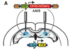
hM3Dq receptor, is a Gq-coupled receptor which responds to CNO resulting in depolarization and spiking in pyramidal neurons. 
Stimulus -----> expression of hM3Dq receptor (in limited to a fraction of excitatory neurons resulting in c-fos promotor drive) -----> So, neurons can be genetically tagged in this way
DOX was used to control in order to capture the receptor expression within a certain time window.
Note: the neural ensemble that got active by this "natural" way, can be reactivated "artificially" again by CNO exposure. This artificially induced activity preserves the spatial character of the neural ensemble. However, it does not preserve the temporal dynamics achieved by natural stimuli (?)
Activity induced by hM3Dq is not temporally coordinated in response to the inducing stimulus (CNO), as is the case with ChR2-driven stimulation by light. 

Experiment design:


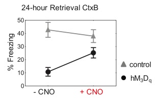
On day 1, mice exposed to CtxA . On day 2, mice got DOX and CNO, then in CtxB got shocked. 24h later tested. 
1) Whether the CtxA neurons synthetic activation served as a conditioned stimulus to produce associative fear memory(?)
If so, mice would freeze to CNO or CtxA itself indicating natural and artificial activation of the neurons were similar  --> was not the case  

Also, CtxA neurons were activated by CNO, and shocked right away in CtxB resulting in no CNO-dependent memory. 
Also, when neurons got tagged during conditioning, CNO did not generate freezing. 
So, the first option is dismissed.

2) Whether the shocking experience in CtxB would take over and mice would show normal conditioning to CtxB (?)
Mice showed severe freezing deficit to CtxB indicating CNO-induced activity interfere with normal encoding memory for CtxB. 

3) A hybrid representation would form as a result of incorporation between CNO-induced artificial stimulation + natural sensory cues from CtxB. (Above) Seeing high freezing to CtxB in CNO+ confirms the third suggestion. 

Note: A correlation between freezing during memory retrieval and the degree of neural activation, as assessed by c-fos expression in the hippocampus.  


Question: Whether retrieval involves activation of some neurons, which were active during the initial learning (?)
Experiment design:

Result indicates: CNO-induced activation of a competing neural network interferes with the learned spatial code and degrades recognition if this activity was not present during the initial training. This is not surprising, given that even limited focal hippocampal stimulation has been shown to disrupt spatial memory: 


Question: To address ensemble specificity ----> Does the hybrid fear memory formed by hM3Dqfos mice incorporate the specific pattern of ctxA neurons activated by CNO during learning, or are the mice responding to a less specific alteration in brain state?


They put mice on DOX after CtxB training to let turnover of the receptor. 2 days later they DOX off and exposure to CtxC. 2 weeks later test.

Result indicates: A requirement for reactivation specifically of the learned (ctxA) neural ensemble, rather than a generalized change in brain state caused by CNO-induced activity.

Supporting design addressing the same above question:  


Result : When mice were fear-conditioned after injection of CNO to artificially activate the ctxB ensemble during learning, they developed wild-type levels of 24-hour context fear memory that were independent of CNO stimulation. This is in contrast to the deficit produced in mice pre-exposed to the novel CtxA and further supports the hypothesis that there must be a match in the spatial pattern of neural activity at learning and retrieval. 

Paper's conclusions: 


- Flexibility in the specific neurons incorporated into a fear memory trace in the amygdala through a selection mechanism in which more excitable neurons are preferentially incorporated into the trace do not appear to be attributable to this type of selection, as the reactivation of the neurons with CNO is required for retrieval, whereas in the previous studies the stimulated neurons were part of a representation that could be naturally retrieved. 
This difference may be due to different requirements for forming simple associations in the amygdala versus more complex representations in the hippocampus and cortex. 


- They found that the CNO activation alone could not act as an independent cue. 

- It is possible that uncoordinated CNO-based stimulus could serve as a conditioned stimulus if it was limited to a discrete primary sen- sory area, such as the piriform cortex (?)

- Although the CNO-basedstimulation does not replicate the temporal dynamics of this naturally occurring internal activity, the approach allows the activation of a distributed spatial pattern of neurons recruited during a specific experience (ctxA exposure). 

- Their results show that this spatial pattern of activity at the time of learning and retrieval must match for appropriate recall. The results imply a strong spatial component to coding in this form of learning and support the idea that the internal dynamics of the brain at the time of learning contribute to memory encoding. 

Friday, April 12, 2013

Do the learning and retrieval of a memory activate the same neurons? Mayford- August 2007

*Dox: Doxycycline is used in "Tet-on" and "Tet-off" tetracycline-controlled transcriptional activation to regulate transgene expression in organisms and cell cultures.
*tau- LacZ (LAC): Marker genes to trace neuronal activity during learning (c-Fos in this paper)
*Zif/Egr (ZIF): An immediate early gene; as a neuronal activity indicator during retrieval
*Teto: A promoter
A transgenic mouse, TetTag: tTA (tetracycline transactivator system) linked to the c-fos promoter. 
Dox was used to preserve the c-Fos+ cells activity during the FC training until the retrieval, since they cut off the DOX to visualize those activated cells- as well as- capturing the ZIF+ cells shortly after the test. 

12% of neurons tagged with LAC were
reactivated during retrieval. 
The non-reactivated cells were not specific 
to the CS-US training. They think the reactivated neurons could be the possible component of a stable engram or memory trace for conditioned fear.
There was no correlation between the estimated freezing during tone retrieval and the number of reactivated neurons in the BLA. However, the number of reactivated neurons in the lateral amygdala (LA) correlated with the estimated freezing during tone retrieval but did not correlate with the freezing during context retrieval